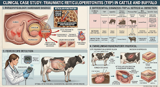
Traumatic reticulo-peritonitis also known as Hardware disease is a critical surgical and medical emergency in adult cattle and buffalo. Animals tend to swallow dense foreign bodies from their indiscriminate and rapid feeding behavior without prior mastication as they do not have tactile discrimination by the oral mucosa. While smooth blunt foreign bodies like stones and plastics remain retained inert in the ruminal vault, sharp linear metallic foreign bodies like wires, nails, needles, and blades sink into the deeper aspect of the reticulum. Reticular contractions drive sharp metallic foreign bodies into the reticulum wall precipitating local peritonitis, diaphragmatic perforation, or traumatic pericarditis.
1. Anatomy and pathophysiology of penetration
The ruminant stomach comprises four compartments namely: Rumen, Reticulum, Omasum, and the Abomasum.
1. The ingested forage is temporarily stored in the rumen for microbial fermentation to produce Volatile Fatty Acids (VFAs).
2. The reticulum, whose honeycomb musculature filters and traps foreign bodies, holds dense metallic foreign bodies at the bottom.
3. Normal reticular contractions force sharp metallic foreign bodies against the reticular wall:
Local TRP : Puncturing of the reticular wall with local peritonitis and fibrinous adhesions.
Traumatic pericarditis: Puncturing of the anterior aspect of the diaphragm into the thoracic cavity and pericardial sac precipitating septic pericarditis, friction rubs, and congestive cardiac failure.
2. Clinical signs and physical examination
Animals with acute or subacute TRP adopt distinctive postures and have specific systemic clinical signs.
Abducted elbow gait and Kyphosis: Animals stand with their elbows abducted outward and the spine arched to reduce pressure on the reticulum.
Reluctance to move: They have severe hesitation to move, turn tightly, walk up or down slopes.
Postural fixation: They have an aversion to lying down or rising and can remain standing or lying still for hours. Pinching the animal along withers provoke grunting, bruxism, or refusal to dip the back due to ventral peritoneal pain.
Progressive weight loss: Anorexia results in emaciation, rough, and dry hide while the body temperature is normal or subnormal in non-septic TRP.
3. Differentiating TRP, Ketosis, and Impaction
Since anorexia, decreased milk yield, and progressive weight loss are features of many bovine metabolic diseases, it is essential to perform a clear differential diagnosis between TRP, Ketosis, and Impaction.
Traumatic Reticulo-peritonitis (TRP): TRP develops after reticular wall puncture by sharp metallic foreign bodies. Affected animals present with abducted elbow gait, kyphosis, and reluctance to move irrespective of the lactation status. Rectal examination reveals scanty hard feces due to ruminal stasis. Characteristic diagnostic features include pain response to xiphoid pressure (Withers pinch test) and presence of metal detected by a Metal Detector or Ferroscope. TRP does not respond to routine fluid therapy and antibiotics but requires an emergency Left Paralumbar Rumenotomy. A positive response to intra-ruminal magnet therapy confirms the diagnosis of Impaction.
Bovine Ketosis (Acetonemia): This condition results from the animal’s negative energy balance (NEB) common in early lactation (25-40 L/day) in cows (1-2 months post-partum). Acetonemia animals have normal gait or exhibit nervous circling behavior while passing small hard fecal balls. The pathognomonic sign of Ketosis is the sweet, fruity acetone odor from breath, milk, and urine. Ketotic animals respond rapidly to IV Dextrose and Glucocorticoids.
Ruminal Impaction: Impaction develops after ingestion of dense fibrous fodder or plastics. Affected animals have swollen abdomen with a lethargic attitude irrespective of their lactation status. Rectal examination reveals no feces in the rectum or dark sticky mucus-coated feces. Characteristic finding is a doughy mass in the left paralumbar fossa palpated on rumen examination. Impacted animals respond to IV fluid therapy and oral mineral oil/laxative administration.
4. Dr.’s clinical thought process: diagnostic verification and triage
🩺 Clinical rationale on TRP management:
“When suspecting TRP in a dairy cow, avoid initial oral therapies for ruminal stasis and scan the reticular area for the presence of any metallic foreign bodies using a Metal or Ferroscope. Avoid testing for metallic foreign bodies when the animal has neck chains or collars by removing them to prevent false positives. If the early stages of TRP (metallic foreign body presence in reticulum) are detected, you can safely resort to the emergency Left Paralumbar Rumenotomy procedure to extract the foreign body and repair the reticular wall. Trying to treat a penetrating wire in TRP by only giving oral magnets will be ineffective. However, if the wire in TRP has already pierced through the diaphragm into the thoracic cavity precipitating septic pericarditis, the prognosis for the surgical procedure becomes very poor due to the altered pressure dynamics in the thoracic cavity during exploratory surgery. The earlier the Left Paralumbar Rumenotomy is performed before the diaphragm is breached, the better is the prognosis.”
5. Surgery: Left Paralumbar Rumenotomy
Pre-surgical preparation
1. Stand the animal.
2. Apply regional anesthesia by a Left inverted L-block or Line block using Lignocaine Hydrochloride 2% (20 to 30 mL).
3. Prep the left paralumbar fossa with Povidone-Iodine and Alcohol.
Surgical procedure
1. Laparotomy Incision: Make a 15 to 20 cm vertical incision over the skin, muscles, and peritoneum at the center of the left paralumbar fossa.
2. Rumen anchoring: Lap-rotate and anchor the rumen wall to the edge of the skin using continuous suturing (or a Weingarth frame) to prevent contamination of the abdominal cavity.
3. Rumenotomy Incision: Make a vertical incision over the rumen wall.
4. Reticular examination: Insert a gloved hand down and forward into the honeycomb reticulum and systematically palpate for any embedded wires, nails, or loose metallic foreign bodies along the ventral floor of the reticulum.
5. Foreign body removal and magnet placement: Carefully pull out any embedded foreign bodies from the reticulum wall back into the rumen lumen and place an oral Rumen Magnet in the reticulum to trap any other metallic foreign bodies.
6. Closure: Close the rumen wall using a double-inverting suture pattern (Lambert/Cushing) with Chromic Catgut No. 2 followed by a standard layered closure of the abdominal cavity.
6. Post-operative medication
Post-operative care involves infection control, analgesia, and restoring normal ruminal motility over an 8 to 15-day healing period.
1. Parenteral antibiotic therapy
Procaine Penicillin & Dihydrostreptomycin: 20,000 IU/kg IM QD for 7 to 10 days
OR
Ceftiofur Sodium: 1.1 to 2.2 mg/kg (0.5 to 1.0 mg/lb) IM/SC QD for 5 to 7 days.
1. Analgesic and anti-inflammatory therapy
Meloxicam:
Dosage (mg/kg): 0.5 mg/kg
Dosage (mg/lb): 0.23 mg/lb
Route: Intramuscular (IM) QD for 3 to 5 days.
Flunixin Meglumine (Alternative):
Dosage (mg/kg): 1.1 to 2.2 mg/kg
Dosage (mg/lb): 0.5 to 1.0 mg/lb
Route: Intravenous (IV) QD for 3 days.
1. Fluids and ruminal motility support
Isotonic fluid therapy: Intravenous (IV) Normal Saline (0.9% NaCl) or Ringer’s Lactate (2 to 4 Liters IV daily).
Rumen microflora transfaunation: Administer 2 to 5 Liters of fresh ruminal fluid and oral yeast supplement to help reestablish VFA production.
Dietary transition: Feed soft, easily digestible green fodder in small frequent meals and gradually resume concentrate rations over 2 weeks.