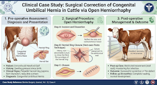
Umbilical hernias can be defined as the most common congenital structural defects or some time due to accident and hitting by another animals encountered in young cattle. They occur due to incomplete closure or delayed contraction of the umbilical ring after birth, allowing abdominal contents-most commonly omentum or loops of intestine-to protrude through the ventral abdominal wall.
By observing physical body conditions; small defects may occasionally go unnoticed, larger umbilical defects pose a continuous risk of enlargement, bowel adhesion, intestinal incarceration or life-threatening strangulation if hernia condition is not properly solved at initial stage. Early surgical repair (herniorrhaphy) provides an excellent prognosis and permanently restores abdominal wall integrity.
1. Case History & Clinical Presentation
A young female cattle was presented with a long-standing history of a distinct swelling in the umbilical region that had been present since birth.
Upon clinical examination, the animal demonstrated:
Ventral Umbilical Mass: A soft, well-defined reversible swelling localized precisely at the umbilical site. This hernial mass can be go inside due to pressure by hand and we can observe hernial ring also by finger palpation at site.
Complete Reducibility: The protruding mass was easily reducible on gentle manual palpation, with abdominal contents slipping back smoothly into the peritoneal cavity.
Palpable Hernial Ring: Digital palpation revealed an enlarged umbilical ring defect capable of easily admitting two to three fingers.
General Health Status: The animal maintained normal posture, systemic vital parameters, appetite and gut motility, confirming an uncomplicated hernia without strangulation.
2. Field Diagnostic Assessment & Triage
Physical and anatomical evaluation confirmed a diagnosis of uncomplicated congenital umbilical hernia. Because the hernial ring was wide enough to allow potential migration and trapping of intestinal loops, elective surgical herniorrhaphy was planned to prevent future incarceration or strangulation.
3. Dr.’s Clinical Perspective
🩺 Clinical Rationale on Bovine Herniorrhaphy:
“Early surgical intervention in congenital umbilical hernias is essential for preventing long-term complications. Operating while the hernia remains uncomplicated and reducible avoids the grave risks associated with intestinal strangulation and tissue necrosis. The core surgical objectives are meticulous dissection of the hernial sac, complete reduction of contents and achieving a secure, tension-free closure of the muscular defect using durable absorbable sutures.”
4. Surgical Protocol & Herniorrhaphy Technique
1. Restraint & Preparation: The animal was properly restrained in dorsal or lateral recumbency. The surgical site around the umbilical region was shaved, thoroughly cleaned and prepared aseptically.
2. Local Anesthesia: Infiltration of local anesthetic solution (2% Lignocaine Hydrochloride) was administered around the planned incision site.
3. Incision & Sac Dissection:
A linear skin incision was carefully performed over the apex of the umbilical swelling.
The underlying hernial sac was meticulously dissected free from the surrounding subcutaneous tissues and abdominal fascia.
4. Reduction & Closure of the Defect:
The hernial contents were carefully evaluated and repositioned into the abdominal cavity.
The fibrous edges of the hernial ring were freshened and closed securely using appropriate suturing techniques with heavy absorbable suture material to eliminate the defect.
5. Tissue Closure & Bandaging:
Subcutaneous tissue and muscle layers were apposed using absorbable sutures to eliminate dead space.
The skin was closed using non-absorbable simple interrupted sutures.
The surgical wound was dressed with an antiseptic solution and protected with a sterile bandage.
5. Post-Operative Care & Recovery Management
Systemic Medication: Post-operative administration included broad-spectrum antibiotics to prevent wound infection and non-steroidal anti-inflammatory drugs (NSAIDs) for pain management and reduction of tissue edema.
Wound Hygiene & Dressing: Daily antiseptic dressing changes were recommended to keep the surgical site clean, dry and protected from environmental contaminants.
Activity & Movement Control: The owner was advised to maintain proper wound hygiene and restrict the animal’s physical movement in a clean pen until complete tissue healing was achieved.
Outcome: The animal responded favorably to surgical correction, achieving full healing and complete restoration of abdominal wall strength without recurrence.